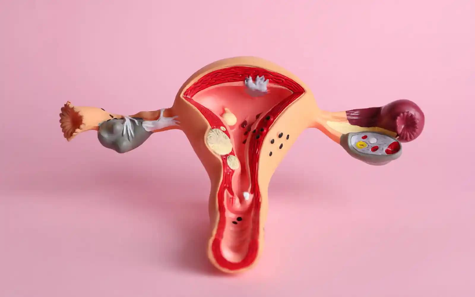
The uterus, Fallopian tubes and ovaries are the crucial organs for women’s fertility. While the ovaries release the eggs and sex hormones which control the function of the other organs, significant events happen in the uterus and Fallopian tubes. Eggs and sperm fertilise in the ends of Fallopian tubes, and embryos grow to 100 cells as they journey to the womb, where they must implant 7-8 days later for pregnancy.
- The Fallopian tubes are delicate but dynamic structures about 10cm (3 inches) long
- They’re essentially extensions of the uterus that form a link to reach the womb eggs released from the ovaries
- Other terms include ‘uterine tubes’, ‘oviducts’, and ‘salpinges’, which can be part of medical words like ‘hydrosalpinges‘

By https://www.scientificanimations.com
Fallopian tube function
-
-
- They link the ovaries and the uterus
- They provide nourishment to sperm, enabling them to survive for up to five days
- The finger-like ends (fimbriae) of tubes ‘sweep’ over ovaries to ‘catch’ eggs after ovulation
- Fertilization usually happens at the end of the tube
- Little hair-like cells called “cilia” in their walls actively propel the embryo to the uterus
- Specialized “peg” cells in the tube walls nourish the growing embryo
- The embryo is transported to the womb after five to six days
-
The uterus function
The womb is an expandable “baby carrier” that has three layers of tissue, which from the inside out are:
- The endometrium lines the inside of the womb and is where an embryo implants
- The myometrium is the smooth muscle that makes up the bulk of the womb
- The perimetrium is the loose connective tissue that surrounds the uterus
The womb is held in position by lots of ligaments. This is usually in a forward position, but sometimes it’s ‘tipped’ backwards in a “retroverted” position. However, the position of the uterus doesn’t affect fertility in any way.

The endometrium
The endometrium lining inside the womb is itself made up of two distinct cell layers with different functions:
- The “functional layer” is the upper layer where an embryo implants and is the layer that’s lost in a period
- The “basal layer” lies under the functional layer and attaches to the muscle of the myometrium. It’s never shed and is the base from which the ‘functional layer’ grows each cycle
The cycle
- The follicular phase relies on estrogen to stimulate the functional layer to grow. The large follicles produce most estrogen in the ovaries. Young women typically have more large follicles than older women, so their womb linings are thicker
- The luteal phase relies on progesterone to transform the functional layer of cells that grew in the follicular phase. Progesterone is produced by the corpus luteum that develops from the follicle that released the egg. The progesterone stimulates the endometrium to become vascular, spongy and sticky to maximize the chances of implantation. The womb surface also has proteins on it for about two days that affect its bonding properties. During two days, called the “window of implantation”, the proteins change and increase the likelihood of attachment
The different phases of the menstrual cycle and the endometrium pattern are:
| Phase | Days | Thickness | Functional epithelium |
|
|---|---|---|---|---|
| Menstruation | 1 – 4 | Thin | 2-4mm | Absent |
| Early follicular | 4 – 10 | Intermediate | 5-7mm | Columnar |
| Late pre-ovulatory | 10 – 14 | Intermediate | Up to 11mm | Columnar |
| Luteal | 15 – 28 | Thick | 7-15mm | Columnar + branches of uterine artery |
The womb lining needs to be 7 – 15mm deep (in the luteal phase) for an embryo to have a reasonable chance of implanting. i ii
The Blood Supply
The ovaries and uterus have to have an adequate blood supply to function and for a pregnancy to progress:
- The flow can be measured by the ‘pulsatility index’ (PI) using a ‘Doppler’ ultrasound scan
- The blood flow varies with changes in sex hormone levels across the cycle
- When the PI is low in the uterine artery, it reduces implantation rates iii
- When the PI rises above 2.5-3.3 in the uterine artery, pregnancy rates fall iv v
- Ultrasound Doppler can identify when (i) pregnancies are at risk of pre-eclampsia, and (ii) babies are small for age (IUGR) due to restricted blood supply vi
Herbal medicine can improve blood flow to the uterus and help foetal growth rates, which is a crucial issue in many pregnancies. vii
ii‘The detrimental effect of increased endometrial thickness on implantation and pregnancy rates and outcome in an in vitro fertilization program’ Ariel Weissman, Lynda Gotlieb, Robert F Casper, Presented in part at the 53rd Annual Meeting of the American Society of Reproductive Medicine, Cincinnati, Ohio, October 18–22, 1997.
iii ‘Ultrasonographic predictors of implantation after assisted reproduction’ Coulam CB, Bustillo M, Soenksen DM, Britten S. FertilSteril 1994;62: 1004 – 1010
iv ‘Doppler assessment of uterine artery blood flow for the prediction of pregnancy after assisted reproduction treatment’ Ultrasound Obstet Gynecol 2008;31: 371 – 375
v‘Uterine and ovarian arteries blood flow during the mid luteal phase in women with unexplained infertility’ M A Razik et al. Middle East Fertility Society Journal Volume 20, Issue 3, September 2015, Pages 209–212
vi ‘Uterine artery Doppler and subendometrial blood flow in patients with unexplained recurrent miscarriage’ H A Wahab et al. Middle East Fertility Society Journal Volume 16, Issue 3, September 2011, Pages 209–214
vii ‘Measuring the effectiveness of Chinese herbal medicine in improving female fertility.’ Trevor Wing and Elke Sedmeier. Journal of Chinese Medicine. 80. Feb. 2006. 22-28
Illustration from Anatomy & Physiology, Connexions Web site. http://cnx.org/content/col11496/1.6/, Jun 19, 2013.