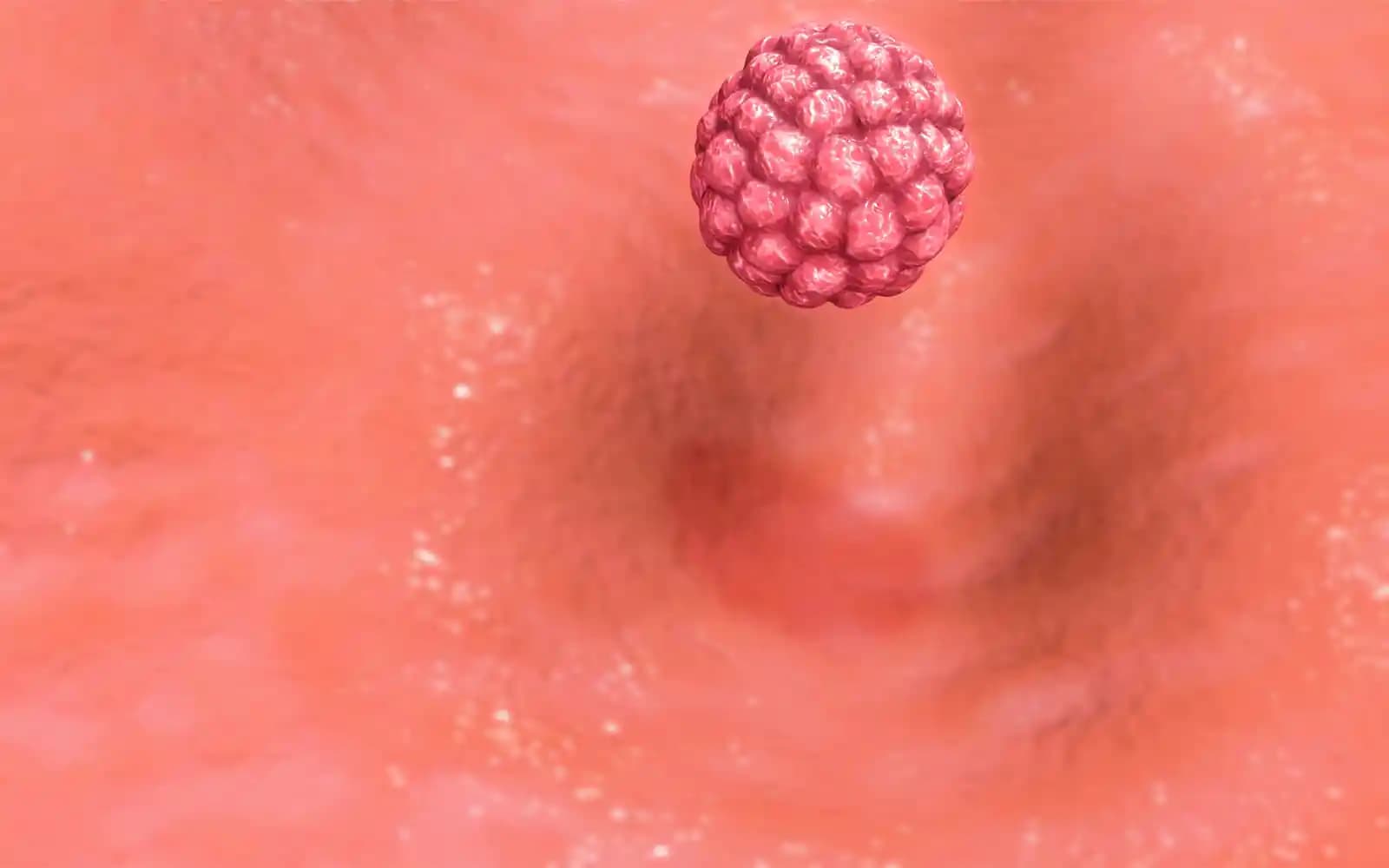
Implantation failure is a significant cause of infertility (and the reason given for about 70% of IVF failures) but is often overlooked as a reason for not conceiving. There are, of course, differences between IVF cycles and natural conceptions and a variety of reasons why healthy embryos don’t become babies. Implantation is a complex process that requires communication and staged connections between an embryo and mother.
IVF has pushed scientific boundaries and extended our knowledge of conception, and it turns out implantation failure is a significant cause of unsuccessful cycles. There is a small percentage of women who experience repeated implantation failure (RIF) with IVF, in which ten or more good-quality embryos don’t succeed over 2-6 IVF cycles (RIF has no formal definition, but this makes it more or less definite). i When couples have three or more unsuccessful cycles, it’s really time for some specialist analysis. ii
Treatment for implantation failure varies with the causes, and we highlight what issues the different Fertility Profiles tend to experience, the testing options, plus research on self-help strategies that seem to work.
14 Reasons for implantation failure
1. Abnormal structure of the endometrial lining
Physical alterations or obstructions of the womb lining are a significant cause of low implantation rates, and they include uterine polyps, fibroids, Asherman’s syndrome, or endometrial hyperplasia. iii It’s important to know these abnormalities are often missed during an initial diagnosis, even when a hysteroscopy or hysterosalpingogram (HSG) is performed. We know this because when women have a second internal examination because of repeated implantation failure or repeated miscarriage, an abnormal structure in the womb is found 25% of the time.
2. The endometrium is too thin
The womb’s lining functional endometrial layer needs to be at least 7mm on the 7th day after ovulation to support implantation. v A lining depth under 8 or 9mm is so thin that embryos have difficulty burrowing into it and implanting properly. As the endometrium growth is stimulated by estrogens released from large follicles in the ovary, this is an issue for women who have few large follicles in their cycles. Thinner womb linings are a natural consequence of lower ovarian reserve or abnormally high follicle losses as fewer follicles reach the ovulation stage each cycle. This is an issue that tends to affect older women or women exposed to toxins the most. However, there are other possible causes:
-
- Long-term use of the oral contraceptive pill thins the endometrium
- Clomid thins the womb lining and increases the risk of ectopic pregnancy, and the higher the dose and the longer Clomid is given, the thinner the endometrium is likely to become. [i] [ii]
- Malnourishment reduces the availability of sufficient nutrients to grow a healthy womb lining
- Over-exercising reduces the availability of nutrients to build the womb lining
- Poor blood circulation in either the ovaries or the uterus reduces the transport of estrogens to the womb
Testing is usually done with ultrasound scans that can measure the thickness of the endometrium.
3. Poor blood flow
The uterus requires a healthy blood supply to support its health and function, and low blood flow can restrict the supply of nutrients and hormones needed to build a healthy womb lining or pregnancy. Blood flow in the uterine arteries needs to be above 30 cm/s (3.0 Pl) for the endometrial lining to grow sufficiently to support implantation, and testing is usually done with a Doppler ultrasound. Women who have repeated miscarriage (RP) have uterine arterial blood flow lower than other women, and it’s particularly bad if they have ANA. [iii] The causes of low uterine blood flow are:
 iv
iv
4. Hydrosalpinges
A hydrosalpinx is a grossly distorted Fallopian tube, and they’re usually caused by sexually transmitted infections which trigger inflammation. The far end of the tube gets blocked, and it fills with a toxic mix of blood and mucus fluids which turn the tube into a “sausage”. This fluid can drain into the uterus and create a hostile environment that makes implantation much less likely when a healthy embryo arrives from the other tube. There are other causes for hydrosalpinx, which are:
- Endometriosis
- Surgery to the tubes or lower abdomen
- IUD contraceptives
- An ectopic pregnancy
- Tuberculosis
5. Endometrial development dysfunction
There are a couple of days in the luteal phase when the endometrium structure changes and creates a “window of implantation”. The proteins on the surface of the womb are different and they encourage bonding between cells, which is crucial for the implantation process. Significant differences in these protein bond changes have been seen between women, and evidence is emerging that women who experience RIF (repeated implantation failure) have relatively “down-regulated” bonding. This contrasts with women who have RM (repeated miscarriages), who have “up-regulation” of bonding that may encourage the implantation of sub-standard embryos and partly explain their subsequent loss. [vi]
The timing of the endometrial changes to coincide with the arrival of the embryo is a crucial issue because bonding is less likely to occur if the womb isn’t at optimal receptivity when the embryo arrives. Endometrial receptivity seems the most significant limiting factor in establishing IVF pregnancies with many gynaecological diseases, and causes include:
- PCOS [vii]
- Endometriosis
- Psychological stress
6. Genetic abnormalities
A high percentage of embryos have abnormal genetic make-ups, and this is by far the common reason for implantation failures and early miscarriages. There are two possible genetic problems which can cause implantation failure:
- Major chromosomal problems (aneuploidy) that cause conditions like Down’s Syndrome
- High DNA fragmentation levels creating excessive disorder in the developing embryo vi
7. Heightened immune response
There are immune conditions such as elevated natural killer cells where one side of the mother’s immune system is too active, and it mistakenly removes embryo cells which either prevent implantation or cause miscarriage. Both sides of the immune system need to respond to pregnancy for it to succeed, but an overly aggressive system will remove any unfamiliar cells, including potential babies.
While this is considered the most common immune-based cause of implantation failure, the “suppressive” side of the immune system is sometimes at fault if it fails to protect the embryo from a normal healthy responses by the “aggressive” side.
8. The parents’ immune systems are too similar
The immune system relies on the ability to distinguish cells that are “self” from “non-self”, and this involves HLA-DQα markers on our cell walls that flag up “self” to the immune system. If the two partners have exactly the same HLA-DQα markers, their embryo will look like “self” to the mother’s immune system, and while this may sound fine, a mother’s immune system has to recognise a pregnancy to be able to make the necessary adaptations that accommodate it.
9. Uneven Endometrium
Every period involves “shedding” of the womb’s functional lining, and if this doesn’t happen across the womb, it leaves an uneven functional layer behind. The functional layer regrows in the follicular phase, and the womb will end up with a mix of old and new tissue on the surface, which is where implantation occurs. The age, depth and health of the functional layer affect the ability to bond with an embryo, and implantation is less likely in old tissue. Many women with this condition experience “dysfunctional uterine bleeding,” which involves heavy periods, clots or periods that vary greatly from month to month, but other causes of an uneven functional endometrial lining are:
- Endometriosis
- Endometrial hyperplasia
- Infection or inflammation of the endometrium
- Endometrial cancer
- Fibroids and polyps
10. Excessive blood clotting
Thrombophilia is a significant issue for women during pregnancy as it increases the risk of miscarriage, but what’s less well-known is that thrombophilia is more common in women with Repeated Implantation Failure (RIF) or “unexplained infertility” than in the general population. vii There is a multitude of causes of thrombophilia, including:
- Genetics such as factor V Leiden and prothrombin G20210A
- Autoimmune conditions like anti-phospholipid syndrome and lupus anticoagulant that are acquired
- Sedentary lifestyles
- Reactions to the drug Heparin can cause Heparin-induced thrombocytopenia(HIT)
- Nephrotic syndrome
- Estrogens in the combined birth pill or HRT increase the risk of thrombosis 2-6x
- Obesity more than doubles the risk of thrombosis
We recommend thrombophilia testing for all women who experience RIF, RM or unexplained infertility.
11. High “aromatase” levels
Aromatase (also called estrogen synthase) is an enzyme involved in estrogen production, specifically the conversion of testosterone into estrogens. Aromatase is attracting a lot of interest recently, and abnormally high aromatase levels in the womb lining are associated with lower implantation rates. viii There are many causes of high aromatase including:
- Psychological stress
- Inappropriate diet
- Endometriosis
- Excess alcohol
- Inflammation and pain
- Type II diabetes
- Medications including some antibiotics and Phenothiazines
- Genetic predisposition [viii]
12. Low “integrin” levels
Integrins are receptors on cell membranes that regulate the attachments between a cell and its surroundings, and they enable cells to make rapid and flexible responses on their surface and act like glue, which is extremely important for implantation.
The crucial integrin appears to be β3 integrin which is vital for clotting and for bonding between cells. Low integrin levels in the lining of the womb are linked to low levels of implantation and IVF success ix, and low integrin levels are thought to be more likely when the woman has endometriosis, low progesterone levels or a hydrosalpinx. The tests for integrin levels require an endometrial biopsy 8-10 days after ovulation and further lab assessment.
13. Hormones used in ART
The hormones used in Assisted Reproductive Techniques (ART) can affect the structure of the endometrium after ovulation. In one study, only 30% of women taking Clomid (clomiphene citrate) had a normal structure to their luteal phase endometrium, compared to 48% of women given human chorionic gonadotropin (hCG) and human menopausal gonadotropin (hMG). x
14. Abnormal uterine microbiome
Abnormal microbiome environments in the womb are thought to contribute to abnormal implantation processes, but traditionally, women with bacterial vaginosis were the only cases investigated. The recent explosion of research into the microbiome has questioned this narrow connection with fertility, and an IVF study of 130 women found significant differences in pregnancy rate between the 28% of women who had abnormal microbiomes, and women whose microbiome was “normal”:
- 9% of women with an abnormal microbiome got pregnant
- 43% of the women with healthy microbiomes got pregnant xi-xii
Tests for implantation failure
-
- Blood tests for hormonal, immune and blood clotting disorders
- Transvaginal ultrasound can detect:
- The thickness and pattern of endometrial development
- Polyps or fibroids that can physically obstruct implantation
- Follicle development and confirmation of the egg’s release from the follicle
- Ultrasound Doppler can measure blood flow in the uterine artery
- Biopsy of the endometrium to:
- Analyse the immune status of the endometrium (e.g. for NK cells CD57)
- Determine preparation for implantation, specifically for the proteins known to increase the likelihood of attachment, including “beta-3 integrin” (a “biomarker test” for this is now available). Biopsies need to be taken within two days of the standard developmental curve to be accurate; however, biopsy dating of the endometrium is controversial and isn’t advised in routine infertility evaluation xiii
5. Chromosomal “karyotype” tests of the partners (including their HLA-DQ match) is a reasonable option for couples with RIF
6. Microbiome testing of the vagina, gut (or semen)
i ’Investigation and current management of recurrent IVF treatment failure in the UK’. Tan BK, Vandekerckhove P, Kennedy R, Keay SD. BJOG. Jun 2005;112(6):773-80.
ii ’Investigation and treatment of repeated implantation failure following IVF-ET’. Margalioth EJ, Ben-Chetrit A, Gal M, Eldar-Geva T Human Reproduction 21(12):3036-43.
iii “Asherman’s syndrome: a critique and current review”. Klein SM, Garcia C-R (1973). Fertility and Sterility 24 (9): 722–735.
iv By BruceBlaus – Own work, CC BY-SA 4.0, https://commons.wikimedia.org/w/index.php?curid=46604060
iv ’Effect of treatment of intrauterine pathologies with office hysteroscopy in patients with recurrent IVF failure’. Demirol A, Gurgan T. Reprod Biomed Online May 2004;8(5):590-94.
v ’Endometrial thickness appears to be a significant factor in embryo implantation in in vitro fertilisation.’ Noyes N, Liu HC, Sultan K, Schattman G, Rosenwaks Z. Hum Reprod Apr 1995;10(4):919-22.
vi’ Chromosome abnormalities identified by comparative genomic hybridisation in embryos from women with repeated implantation failure.’ Voullaire L, Wilton L, McBain J, Callaghan T, Williamson R. Mol Hum Reprod Nov 2002;8(11): 1035-41.
vii ’Increased rates of thrombophilia in women with repeated IVF failures’. Azem F, Many A, Yovel I, Amit A, Lessing JB, Kupferminc MJ. Human Reproduction February 1, 2004;19(2): 368-70.
viii ’High endometrial aromatase P450 mRNA expression is associated with poor IVF outcome’ Jan Brosens et al. Hum. Reprod. (2004) 19 (2): 352-356.
ix ’Endometrial receptivity defects during IVF cycles with and without letrozole’ Paul B. Miller et al. Hum. Reprod. (2012) 27 (3): 881-888.
x “In vitro fertilisation: the degree of endometrial insufficiency varies with the type of ovarian stimulation”. Sterzik K. et al. University of Ulm, Department of Obstetrics and Gynaecology, West Germany. Fertility and Sterility [1988, 50(3):457-62]
xi Implantation failure of endometrial origin: what is new? Bellver, José; Simón, Carlos. Current Opinion in Obstetrics and Gynecology: August 2018 – Volume 30 – Issue 4 – p 229–236
xii Abnormal vaginal microbiota may be associated with poor reproductive outcomes: a prospective study in IVF patients. Haahr T et al. Hum Reprod. 2016 Apr;31(4):795-803. doi: 10.1093/humrep/dew026.
xiii ’Histological dating of timed endometrial biopsy tissue is not related to fertility status.’ Coutifaris C et al. Fertil Steril. 2004 Nov;82(5):1264-72.
xiv ’Endometrial receptivity defects during IVF cycles with and without letrozole’ Paul B. Miller et al. Hum. Reprod. (2012) 27 (3): 881-888.
xv ’Surgical treatment for tubal disease in women due to undergo in vitro fertilisation.’ Johnson N, van Voorst S, Sowter MC, Strandell A, Mol BW. Cochrane Database Syst Rev (1): CD002125.
xvi “Endometrial growth and uterine blood flow: a pilot study for improving endometrial thickness in the patients with a thin endometrium”. Takasaki A, Tamura H, Miwa I, Taketani T, Shimamura K, Sugino N (April 2010). Fertil. Steril. 93 (6): 1851–8.
xvii ’Effects of ascorbic acid supplementation on serum progesterone levels in patients with a luteal phase defect.’ Henmi H, et al. Fertility and Sterility (2003;80:459–61)
[i] Effects of clomiphene citrate on the endometrial thickness and echogenic pattern of the endometrium.’ Yasuhiko Nakamura M.D. Et al. Fertility &Sterility Vol. 67, No. 2, February 1997
[ii] ‘Clomiphene citrate causes aberrant tubal apoptosis and estrogen receptor activation in rat fallopian tube: implications for tubal ectopic pregnancy’ R Shao, Et al. Biology of Reproduction 80(6):1262-1271. 2009
[iii] T. Habara et al. Elevated blood flow resistance in uterine arteries of women with unexplained recurrent pregnancy loss. January 2002 Human Reproduction 17(1):190-4
[iv] Helbig, Anne et al. “Does antenatal maternal psychological distress affect placental circulation in the third trimester?.” PloS one vol. 8,2 (2013): e57071. doi:10.1371/journal.pone.0057071
[v] Kathirvel Gopalakrishnan et al. Elevated Testosterone Reduces Uterine Blood Flow, Spiral Artery Elongation, and Placental Oxygenation in Pregnant Rats. March 2016. Hypertension. Vol. 67, Issue 3
[vi]</sup Huang, J., Qin, H., Yang, Y., Chen, X., Zhang, J., Laird, S., Wang, C. C., Chan, T. F., & Li, T. C. (2017). A comparison of transcriptomic profiles in endometrium during window of implantation between women with unexplained recurrent implantation failure and recurrent miscarriage, Reproduction, 153(6), 749-758.
[vii] Ione Lopes, et al. Endométrio na janela de implantação em mulheres com síndrome dos ovários policísticos,
Revista da Associação Médica Brasileira, Volume 57, Issue 6, 2011, Pages 702-709,
[viii] CYP19A1 gene: MedlinePlus Genetics